
Cancer care depends on clear information. Patients need answers, care teams need useful images, and everyone benefits when doctors can see what happens inside the body without adding unnecessary stress. Doppler ultrasound helps fill that role in many care settings.
Most people connect ultrasound with pregnancy, but medical teams use ultrasound for many other reasons. In cancer care, ultrasound can help doctors look at soft tissue, examine suspicious areas and guide certain procedures. Doppler technology adds another layer by showing how blood moves through vessels.
That blood flow information can help doctors understand a tumor, plan the next step and monitor changes during care. It doesn’t replace every imaging tool, and it doesn’t answer every question by itself. Still, Doppler ultrasound supports cancer care and provides valuable details in a quick, noninvasive way.
Traditional ultrasound creates images of structures inside the body. It can help a doctor see the shape, size and location of a mass. Doppler ultrasound goes further because it tracks movement. It shows how blood travels through arteries, veins and smaller vessels near an area of concern.
Cancer care teams pay close attention to blood flow because tumors can develop their own blood supply. A tumor may need blood vessels to deliver oxygen and nutrients. When doctors look at that activity, they gain more context about what they see on a standard image.
Doppler doesn’t diagnose cancer on its own. A doctor may still recommend lab work, a biopsy, MRI, CT or another test. However, Doppler can add helpful information while the care team builds a complete picture.
Doctors often use the word vascularity when they talk about blood supply in tissue. A mass with more blood flow may raise different questions than a mass with little or no blood flow. Doppler ultrasound helps doctors view that activity in real time.
This view can help care teams evaluate suspicious areas in the breast, thyroid, liver, kidneys, lymph nodes and other parts of the body. The exam may show blood moving around a mass, through a mass or away from nearby tissue. Those patterns can help doctors decide whether they need more testing.
Dopplers can monitor tumor blood flow during different points in cancer care. A doctor may compare current images with earlier images to look for changes in blood flow, size or structure. Those comparisons can support better conversations about treatment progress.
The diagnostic process can feel overwhelming, especially when someone first hears that a scan found a mass. Doppler ultrasound can help doctors gather more information before they recommend the next step.
For example, a care team may use ultrasound to tell whether a lump looks solid or fluid-filled. Doppler can then show whether blood flows within or around that lump. A fluid-filled cyst often leads to a different care path than a solid mass with active blood flow.
Doctors may also use Doppler when they examine swollen lymph nodes. Blood flow patterns can help them decide whether a lymph node looks reactive, suspicious or worth a closer look. That information can guide timing, follow-up imaging or biopsy planning.
Many cancer diagnoses require a tissue sample. During an ultrasound-guided biopsy, the clinician uses images to place the needle with precision. Doppler can help the clinician see nearby vessels and choose a safer path.
That added detail can reduce guesswork during the procedure. The clinician can avoid blood vessels when possible and target the area that needs sampling. A more accurate sample can help the pathologist evaluate the tissue and help the care team move forward with the right plan.
Patients often appreciate that ultrasound-guided procedures usually happen without radiation. The exam room may still feel intimidating, but the imaging method itself uses sound waves. That feature can make ultrasound useful for repeated checks when the care plan calls for ongoing monitoring.
Cancer treatment often involves careful tracking. Doctors want to know whether a tumor shrinks, grows or changes in character. Size tells part of the story, but blood flow can add another useful layer.
Some treatments aim to disrupt a tumor’s ability to grow. When blood flow changes, doctors may gain early clues about how the tumor responds. A tumor may show less vascular activity, a different flow pattern or changes around its edges.
Doppler ultrasound can help care teams compare images over time. These comparisons can support decisions about whether to continue a treatment, adjust the plan or order another imaging test. The doctor considers the full clinical picture, not one scan alone.
Cancer care already places a heavy load on patients and families. Long appointments, tests and waiting periods can wear people down. Doppler ultrasound can help because the exam usually happens quickly, and many patients return to normal activities afterward.
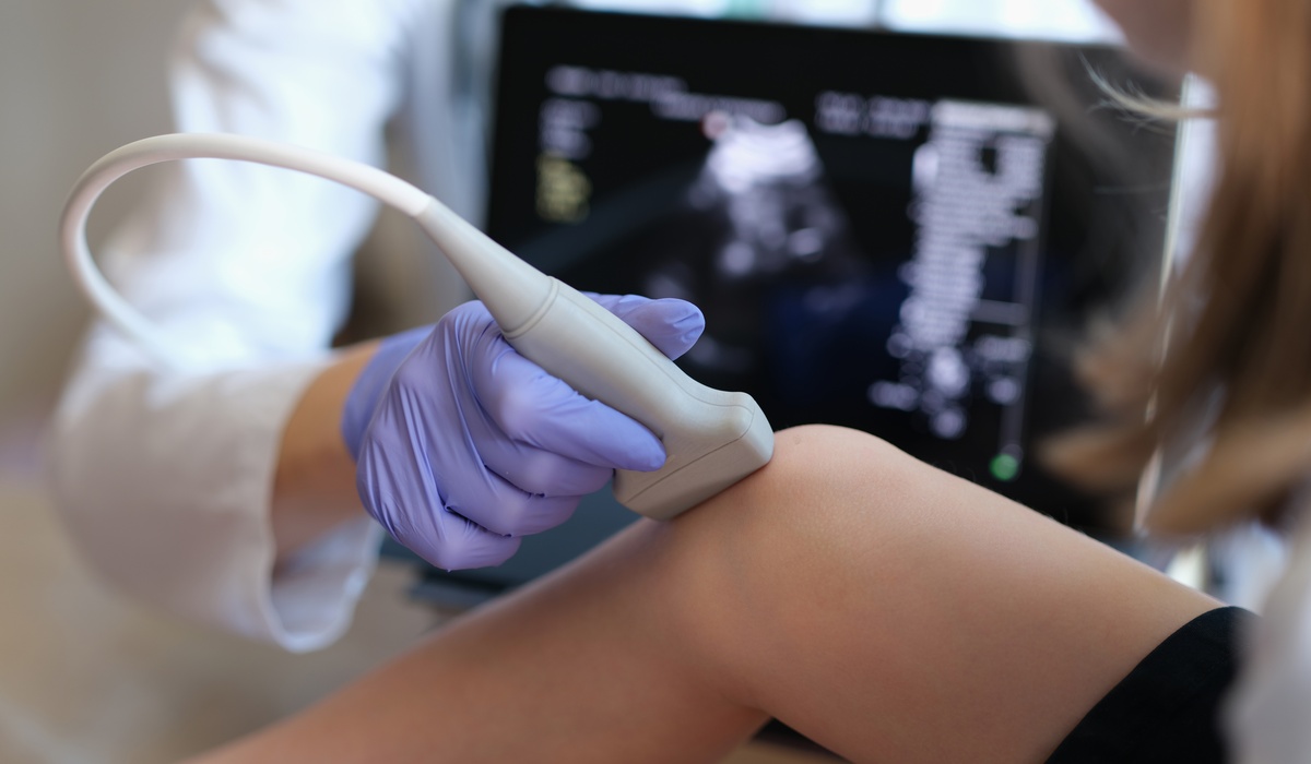
The test doesn’t require an incision. A sonographer or clinician places gel on the skin and moves a handheld transducer over the area. The screen displays images while the device captures blood flow information.
Some exams require preparation, depending on the body area. A patient may need to fast before an abdominal ultrasound, for example. The care team gives instructions before the appointment, and patients should follow those directions closely.
Many imaging tools play important roles in cancer care. CT, PET and X-ray exams can provide essential details, but they use radiation. Ultrasound uses sound waves instead.
That difference can make Doppler ultrasound helpful when doctors need repeated imaging. A patient may need several checks during treatment or follow-up care. Ultrasound can support those visits without adding radiation exposure.
That doesn’t mean ultrasound works for every situation. Sound waves don’t travel well through bone or air, and some tumors sit too deep or in areas that another imaging method can show better. Doctors choose the imaging tool that best fits the question they need to answer.
Good cancer care depends on teamwork. Surgeons, oncologists, radiologists, nurses and primary care providers may all play a role. Doppler ultrasound can give that team shared information that supports clearer communication.
Images can help the care team explain what they see. A doctor may show where blood enters a mass or how a lymph node looks on the screen. Patients often feel more grounded when they can connect a discussion to a visual image.
That information can also help patients ask better questions. They may ask why the doctor recommends a biopsy, why follow-up imaging makes sense or why the team wants a different scan. Clear information can reduce confusion during an already stressful time.
Cancer care doesn’t end when active treatment ends. Many patients continue follow-up appointments for months or years. During those visits, doctors look for healing, recurrence, treatment effects and new concerns.
Doppler ultrasound can help evaluate areas that need attention after surgery, chemotherapy, radiation or targeted treatment. For example, a doctor may use Doppler to check blood flow near a treated area or evaluate a new lump. The exam can help the team decide whether the finding looks stable or needs more testing.
Cancer care moves one decision at a time. Each test should help answer a meaningful question, and Doppler ultrasound can provide valuable insight during cancer care when doctors need to understand blood flow. It can support diagnosis, guide biopsies, monitor treatment response and help with follow-up care.
Patients deserve tools that give clear information without adding unnecessary burden. Doppler ultrasound offers a practical, noninvasive way to gather details that can shape the next step. When doctors use it alongside other clinical information, it can support more confident conversations and more informed care.
About The Author